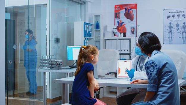
Your heart is the key organ of your circulatory system, which is a system of blood vessels that transport blood throughout your body. It also collaborates with other systems in the body to regulate your blood pressure and heart rate. It is enclosed by your rib cage and is located in your thorax amid your lungs, mostly to the left of the centre. Your heart is around the size of your closed fist and weighs approximately 300 g. The cardiovascular system is made up of the heart, blood, arteries, capillaries, and veins. Your genetic factors, individual medical history, and lifestyle all have an impact on how effectively your heart functions.
Recommended: Salt Lake City Personal Injury Lawyers
The heart is divided into four chambers. The atria are the heart’s two upper compartments which receive blood. The ventricles are the lower two chambers that pump out blood. The septum is a muscular wall that divides the left and right atria and the left and right ventricles. Valves separate the atria and ventricles. The heart’s walls are made up of three layers of tissue. Myocardium: This is the heart’s muscular tissue. Endocardium: the tissue that covers the interior of the heart and supports the valves and chambers. The pericardium is a thin protective layer that covers the other parts of the heart. Epicardium: The inner layer of the pericardium is composed of this protective layer, which is largely made up of connective tissue.
The superior and inferior vena cava are the body’s largest vessels. These veins transport deoxygenated blood from the body to the right atrium. The blood is then sent to the right ventricle whenever the right atrium contracts. The right ventricle will then contract and pump blood via the pulmonary artery to the lungs after getting blood from the right atrium. The blood gets oxygen and releases carbon dioxide here. Once oxygenated, blood will proceed to the heart, specifically the left atrium, through the pulmonary vein. The atrium subsequently contracts, delivering blood to the left ventricle. After receiving all of the blood from the left atrium, the left ventricle contracts and distributes it to the body via the aorta.
The atria and ventricles operate together to pump blood from your heart to other systems by contracting and relaxing alternately. The electrical system of the heart is the source of energy that allows this to happen. Electrical impulses pass through a particular channel throughout the heart muscles to generate your heart rhythm. The impulse originates in the right atrium, in a compact bundle of particular cells known as the SA node (sinoatrial node).
This node is referred to as the biological pacemaker of the heart. The electrical impulses travel through the atria’s walls, causing them to contract. The atrioventricular node is a bundle of cells at the base of the heart that functions as a gate, delaying electrical impulses before they reach the ventricles. This pause allows the atria to contract first before the ventricles. The His-Purkinje system is a combination of fibers that delivers impulses to the ventricle’s muscular walls, causing them to contract.
A typical heart beats at about 50 to 90 times per minute while at rest. Signals from your neurological system and hormones from your endocrine system can regulate how fast and hard your heart is beating. These signals and hormones enable your body to adjust itself depending on the quantity of oxygen and nutrients required. When you workout, for instance, your muscles require more oxygen, causing your heart to beat faster. When you are sleeping, your heart rate will slow down. Excitement, stress, infections, and some drugs also can cause the heart to beat rapidly, occasionally exceeding 100 beats per minute.
Do take your Covid 19 vaccination.
